A veterinary ultrasound uses sound waves to create live images of structures inside your pet’s body. For a dog or cat, it can help a veterinarian examine abdominal organs, the heart, and other soft tissues without surgery, though it does not answer every medical question by itself. If you are wondering “veterinary ultrasound for dogs what does it show,” the short answer is that it can reveal changes in organ size, shape, texture, movement, and blood flow that may help explain a pet’s symptoms.
At Animal Hospital of Rolling Meadows, ultrasound is one of the diagnostic tools veterinarians may use alongside a physical examination, lab work, and other imaging. This guide explains what a scan can show, what the appointment may involve, and how a veterinarian uses the findings to plan next steps. It offers general information, not a diagnosis for an individual pet.
What is a veterinary ultrasound and how does it work?
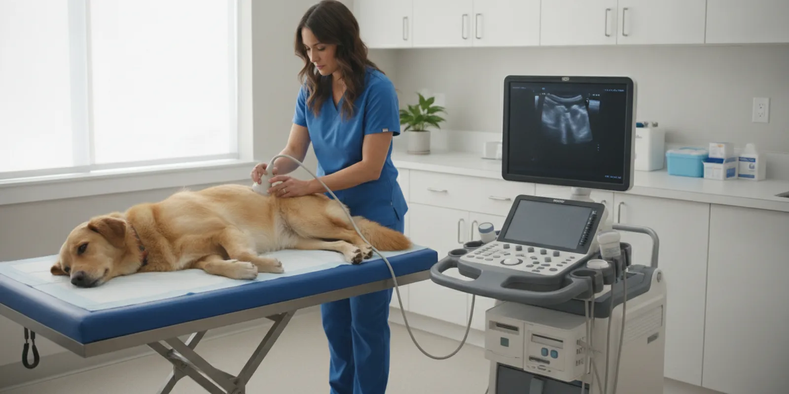
A veterinary ultrasound, also called sonography, uses high-frequency sound waves to produce images of organs and tissues inside an animal’s body. The veterinarian or trained team member moves a small handheld probe over the skin. The probe sends sound waves into the body and receives echoes as those waves bounce back from different tissues. A computer turns the returning echoes into a moving image on a screen.
Unlike an X-ray image, which is especially useful for looking at bones and certain chest or abdominal patterns, ultrasound can show soft tissues in motion. A veterinarian can observe features such as the outline of an organ, the appearance of its internal tissue, and—in some types of examination—blood moving through vessels. The view is produced in real time, so the person scanning can change the probe angle and examine an area from different directions.
Ultrasound is not a single all-purpose test. The question being investigated determines which area is scanned and what kind of examination is appropriate. Common examples include:
- Abdominal ultrasound: A closer look at organs such as the liver, spleen, kidneys, bladder, stomach, intestines, and nearby structures.
- Echocardiography: An ultrasound examination focused on the heart, its chambers, valves, and movement.
- Focused ultrasound: A brief, targeted scan used in certain urgent situations to look for findings such as fluid in body spaces.
- Ultrasound-guided sampling: In selected cases, imaging may help a veterinarian guide a needle to collect a sample. Whether this is suitable depends on the location, the pet, and the clinical question.
Rolling Meadows Animal Hospital lists ultrasound as an in-house diagnostic service. Your veterinarian can explain whether an abdominal scan, heart study, focused scan, or another test best fits your pet’s needs.
What conditions can an ultrasound detect in pets?
Ultrasound can help identify or further investigate changes in soft tissues. It may show that an organ looks enlarged or has an unusual texture, reveal some masses or fluid-filled areas, and help locate certain stones. It may also help evaluate the heart or guide a sample collection. These findings can point toward possible explanations, but an image alone does not always identify the exact cause of a problem.
For example, an abdominal scan may help a veterinarian assess:
- Changes to abdominal organs: The scan may show differences in the size, shape, or internal appearance of the liver, spleen, kidneys, or other organs.
- Some urinary tract concerns: The bladder and kidneys can be examined for certain stones, unusual contents, or structural changes.
- Fluid or masses: Ultrasound can help locate fluid in the abdomen and identify areas that may need closer evaluation or sampling.
- Digestive and pancreatic concerns: The pancreas and gastrointestinal structures may be examined when symptoms and other findings make imaging useful.
- Blood vessels and blood flow: Specialized ultrasound techniques can help assess circulation or investigate certain vascular abnormalities.
- Pregnancy-related questions: Depending on timing and the clinical question, ultrasound may be used to assess pregnancy.
The University of California, Davis School of Veterinary Medicine describes ultrasound as a tool veterinarians use to help diagnose conditions including pancreatitis, enlarged organs, kidney or bladder stones, and portosystemic shunts. The scan helps provide evidence; the veterinarian interprets it in light of the pet’s history, examination, and other test results. Learn more about small-animal ultrasound from UC Davis Veterinary Medicine.
Some problems do not create a clear ultrasound finding, and some findings are not specific to one disease. For example, a change in an organ’s appearance may have more than one possible explanation. A veterinarian may recommend blood or urine testing, X-rays, a sample, a specialist’s interpretation, or monitoring over time to clarify what an image means.
Ultrasound is also not a replacement for every other type of imaging. It may not show detail through gas-filled areas or bone as clearly as other methods, and the quality of the view can depend on the patient’s body, the area being examined, and the equipment and operator. The veterinarian chooses a test based on the question that needs answering.
Ultrasound and other common diagnostic tools
Different tests answer different questions. A veterinarian may use one test or combine several. This comparison describes general strengths, not a recommendation for a particular pet.
| Test | What it can help assess | Important limitation |
|---|---|---|
| Ultrasound | Soft-tissue structures, organ appearance, movement, and some blood flow | Some areas are difficult to view through gas or bone; an image may not identify a cause on its own |
| X-ray (radiography) | Bones, some chest and abdominal patterns, and the size or position of certain structures | It is a still image and may provide less detail of some soft tissues than ultrasound |
| Blood or urine testing | Changes in organ function, blood cells, urine, or other measurable markers | Tests measure different things from imaging and may not show the location or appearance of a structural change |
| CT or MRI | Detailed cross-sectional images for selected clinical questions | May involve referral, specialized equipment, and additional planning; not needed for every case |
Is ultrasound safe for dogs and cats?
Diagnostic ultrasound uses sound waves rather than ionizing radiation, and it is generally considered a noninvasive imaging method. Many routine scans are performed while a pet is awake. The veterinarian will decide how to make the examination comfortable and whether any additional support, such as calming medication or sedation, is appropriate for that individual animal.
“Generally considered safe” does not mean every scan is identical or that no preparation is ever needed. A pet’s health, stress level, ability to remain still, and the reason for the examination all matter. If a procedure such as ultrasound-guided sampling is being considered, the veterinarian will explain the separate steps, possible risks, and alternatives before proceeding.
Ask the veterinary team to explain the plan if you have concerns. Useful questions include:
- Which part of my pet’s body will be scanned, and what question are we trying to answer?
- Will my pet need to fast or have any other preparation?
- Will my pet likely remain awake, and what options are available if they are anxious?
- Could the scan involve a sample or another procedure, or is imaging the only planned step?
- Who will review the images, and when should I expect an update?
Never withhold food, water, or prescribed medication unless your veterinary team specifically tells you to do so. Preparation instructions can vary with the scan and the pet’s medical situation. If a pet has a condition that makes fasting or medication changes a concern, mention it when the appointment is arranged.
What to expect when your pet has an ultrasound
The exact appointment depends on the scan and your pet’s condition. For a planned abdominal examination, the team may first review the reason for imaging, the pet’s history, and any recent bloodwork or X-rays. If preparation is needed, the hospital will give instructions in advance.
During many abdominal scans, the team clips a small area of fur so the probe can make good contact with the skin. A clear gel is applied, and the probe is moved over the area being examined. Your pet may rest on a padded table, and a staff member may gently help them stay in a comfortable position. The scan itself is not usually painful, although holding a sore or anxious pet in a position can be uncomfortable. Let the team know about pain, mobility challenges, or stress before the examination begins.
For a heart ultrasound, the positioning and areas examined are different. The veterinary team will explain what they plan to do and whether the pet’s coat needs any preparation. If the scan is focused on an urgent question, the procedure may be shorter and more targeted than a full diagnostic study.
What you can do to help:
- Share the full story. Describe what you have noticed, when it began, and whether it is changing. Bring a list of medicines and relevant records if they are not already at the hospital.
- Follow only the instructions provided. Confirm food, water, and medication guidance with the veterinary team rather than assuming your pet should fast.
- Tell the team what helps your pet. Mention handling preferences, fear triggers, or mobility needs. Calm, gentle handling can make the visit easier.
- Ask about the timing of results. Some findings may be discussed during the visit; other images may need a veterinarian’s review or an outside radiologist’s interpretation.
- Make a plan for follow-up. Before leaving, confirm who will contact you, what the next step may be, and whom to call if your pet’s condition changes.
At Animal Hospital of Rolling Meadows, ultrasound is part of a broader diagnostic approach. The team also lists veterinary medicine services and other services for dogs and cats. Depending on the concern, an examination, lab testing, or additional imaging may be recommended along with ultrasound.
If symptoms are worsening or you are unsure how quickly your pet needs to be seen, contact a veterinary team for guidance. You can read about the hospital’s urgent care services in Rolling Meadows. For general context on signs that may warrant attention, see this guide to signs of pain in dogs. Neither a web guide nor an imaging test replaces an examination.
How ultrasound results guide treatment decisions
An ultrasound result is one part of a clinical picture. The veterinarian considers the images together with your pet’s symptoms, physical examination, age and medical history, and any test results already available. The scan may help narrow the possibilities, indicate that another test is needed, or provide information for choosing a treatment or monitoring plan.
After the scan, a veterinarian may discuss one of several paths:
- The images help answer the question. If the findings fit the symptoms and other test results, the veterinarian can explain what they mean and discuss appropriate care.
- More information is needed. A blood or urine test, X-ray, sample, follow-up scan, or specialist review may help clarify an uncertain finding.
- A finding needs monitoring. In some situations, the veterinarian may recommend watching for changes and rechecking at a suitable time.
- A procedure or referral should be considered. If imaging identifies a concern that needs further treatment or expertise, the veterinarian can discuss options and whether referral is appropriate.
Ask the veterinarian to explain the result in plain language. You might ask what was seen, what could not be assessed, how confident the team is about the interpretation, and how the finding relates to your pet’s signs. If there are several options, ask what each is intended to accomplish and what follow-up is involved.
In some cases, ultrasound helps guide a needle to obtain a cell or fluid sample. A sample can provide information that imaging alone cannot, but sampling is not appropriate for every area or every patient. The veterinarian weighs the possible value and risks, explains the procedure, and discusses alternatives before recommending it.
When a pet needs care beyond diagnostic testing, the next step depends on the findings and the animal’s overall health. Animal Hospital of Rolling Meadows provides a range of care, including surgical services; a scan does not automatically mean surgery is needed. The goal is to use the evidence available to make a reasoned, individualized plan with you.
For a broader look at the practice and its team, you can visit About Animal Hospital of Rolling Meadows or learn more about the veterinary staff. Bring any questions about your pet’s specific results to the veterinarian who examined them.
Frequently asked questions
Does an ultrasound show everything inside my dog?
No. Ultrasound provides useful images of many soft tissues, but it cannot answer every question or show every area equally well. Gas, bone, the location of a structure, and the pet’s movement can affect the view. The veterinarian may recommend another test if the scan does not provide enough information.
Will my dog or cat need to be sedated?
Many pets have routine ultrasound scans while awake, but the decision depends on the type of scan, the pet’s comfort and ability to remain still, and their medical condition. The veterinary team will explain the plan and discuss whether calming medication or sedation is appropriate.
Does my pet need to fast before an ultrasound?
Preparation varies with the examination and your pet’s needs. Ask the hospital for instructions before the appointment, and do not change food, water, or medication routines unless the veterinary team tells you to.
Can an ultrasound diagnose cancer?
An ultrasound may identify a mass or other abnormal area, but the image alone may not determine whether it is cancer. A veterinarian may recommend a sample or additional testing to learn more. The appropriate next step depends on the finding and your pet’s overall situation.
How soon will I know the results?
Timing depends on the examination and whether the images need additional review. Ask the veterinary team who will explain the findings and when you should expect to hear from them. If your pet’s condition changes while you wait, contact a veterinarian promptly.
Ultrasound can give your veterinary team a closer look at organs and soft tissues, but its value comes from interpreting the images alongside your pet’s signs and medical history. If your dog or cat may need an ultrasound, Animal Hospital of Rolling Meadows can help discuss the appropriate diagnostic steps for that individual pet.